
Patna: The city's Paras HMRI Hospital has given a new lease of life to two arrhythmia patients using 3D electro-ana-tomical mapping. Arrhythmia is a condition in which one suffers from irregular heartbeats.
The 3D electro-anatomical mapping allows cardiologists to see the inside of a heart in 3D format. This makes diagnosis accurate. The hospital recently hired a 3D mapping machine from a Calcutta-based facility to treat two patients. It had to pay around Rs 1 lakh to hire the machine.
According to Paras officials, this was for the first time a hi-end diagnostic tool was used for a child in Bihar or Jharkhand.
One of the two patients on whom 3D electroanatomical mapping was done was a 10-year-old boy and the other patient a 18-year-old girl who recently took admission into BA and was skipping college because of health complications. The girl's heartbeats had reached 180 beats per minute against the normal of 60-100 beats per minute.
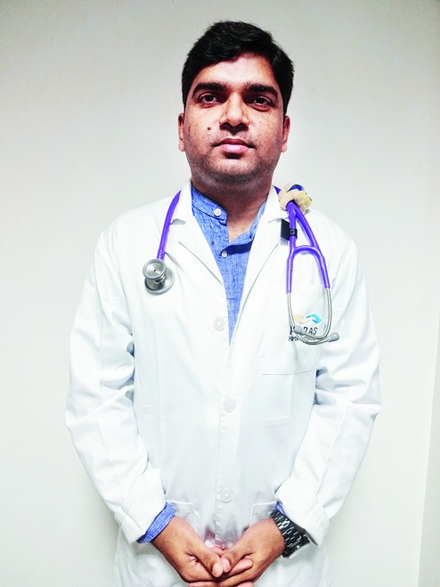
"You can see the heart anatomy in 3D with the machine we have bought," said electrophysiology specialist Narendra Kumar at Paras HMRI Hospital.
"In the traditional method, doctors rely on a 2D image of the heart which he/she gets through X-ray but the heart has a 3-dimensional anatomy. So, getting 3D image of the heart will ensure accurate treatment as doctors will be able to diagnose complex arrhythmia problems with it. With the help of 3D electro-anatomical mapping, we got to see 3D images of the heart and could locate the extra wire in a heart, which was burnt to rid patients of the disorder.
"Basically, we performed cardiac ablation after diagnosing the patients' condition through 3 D electro-anaomical mapping. Cardiac ablation is a procedure to destroy tissue in heart that's allowing incorrect electrical signals to cause an abnormal heart rhythm."
Dr Narendra conducted the 3D electro-anatomical mapping on the patients before cardiac ablation. "As far as our information goes, this is for the first time that this 3D electro-anatomical mapping has been used on any child patient in Bihar and Jharkhand," he said.
He added that the hospital pooled the treatment cost from outside for the two patients from economically weaker sections.
"The 3D mapping machine is available at very few places in the country. If we need to do this procedure again at Paras, we would have to hire it from outside," said Narendra, who was earlier associated with a Netherlands-based hospital for seven years.