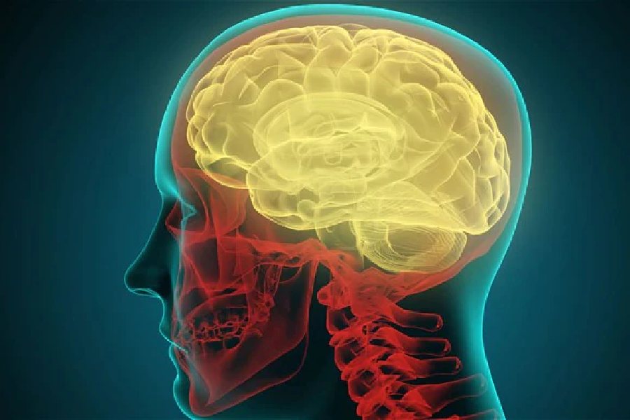
Scientists have created a 3D reconstruction of a millimetre-sized fragment of human brain tissue, about half the size of a rice grain, covering in vivid detail over 57,000 cells and 150 million neural connections, represented through 1,400 terabytes of data.
The work by scientists at Harvard University and Google Research represents a landmark advance towards gaining insights into the physical mechanisms that underpin the normal and disordered human brain. Their paper describing their study was published in the US research journal Science on Thursday.
Despite great advances in neuroscience in recent decades, detailed knowledge about the neural circuitry underlying human brain functions is lacking. Neuroscientists still know little about how the brain’s cellular microstructure and the neural connections support normal brain functions or are disrupted in brain disorders.
In the new study, the researchers relied on a 1 cubic mm fragment of brain tissue removed from a 45-year-old woman during surgery to gain access to an epileptic focus in the underlying hippocampus.
They used electron microscope images and artificial intelligence to colour-code and reconstruct the brain tissue in 3D.
Their reconstruction contains roughly 57,000 cells, about 230 millimetres of blood vessels, and nearly 150 million synapses, making up 1,400 terabytes of data, or equivalent to over 2,80,000 two-hour movies.
Jeff Lichtman, professor of molecular and cellular biology who led the Harvard team, specialises in the field of connectomics, analogous to genomics but seeks to create a comprehensive catalogues of brain structure down to individual cells and their connections with other cells.
“Just a miniscule teeny-weeny bit of the human brain is still thousands of terabytes,” Lichtman said in a media release. The Harvard-Google team has developed a suite of publicly available tools that researchers across the world could use to annotate the connectome.
“Given the enormous investment put into this project, it was important to present the results in a way that anybody else can now go and benefit from them,” Viren Jain, a collaborator at Google Research said in the media release.
A scientist, who was not associated with the study, said she was impressed by the level and depth of detail and the reconstruction achieved. “Finally at least a tiny piece of the most complex part of the brain — the cerebral cortex — has been reconstructed,” said Sandhya Koushika, a neurobiologist at the Tata Institute of Fundamental Research (TIFR), Mumbai. “Will it provide us insights? The answer seems to be: yes. Clearly, computational methods married to the old electron microscopy imaging have the ability to give a map of how the brain is connected in unprecedented levels of detail.”