 |
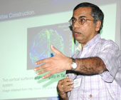
| Prof. Baba C Vemuri at the ISI seminar (Picture by Sanat K. Sinha) |
To track the threats from a vicious tumour or a suicide bomber, scientists have devised a host of sophisticated techniques like image registration, 3D shape and structure analysis, motion and video analysis. ?Image registration is a technique in which two images of different qualities are fused, superimposed, matched or merged to achieve high resolution and finer details,? said Prof. Baba C. Vemuri from the University of Florida, US, while delivering a speech on ?Image Registration with Applications to Medical Imaging? at a recently-held conference on computer vision, graphics and image-processing.
The conference was organised by the Indian Statistical Institute and Indian Unit for Pattern Recognition and Artificial Intelligence. ?Image registration has already proved to be a handy tool in the realm of medical diagnosis,? Vemuri said, arguing that a single medical image is not enough to yield the required information for a flawless diagnosis. ?For instance, single-photon emission computed tomography (SPECT) and positron emission tomography (PET) captures functional body images that only provide physiological information,? said Vemuri. ?On the other hand, computed tomography (CT) and magnetic resonance images (MRI) provide structural images that give an anatomical map of the body.?
According to him, none of the medical imaging techniques is self-sufficient to pin down an elusive tumour. ?This problem is overcome using image registration that merges CT scan and MRI with SPECT and PET, providing physiological as well as anatomical details of a particular section of the body,? Vemuri explained.
Besides image registration, experts are also making use of an image-processing technique called rincipal component analysis (PCA). In PCA, the superfluous part of an image is removed, yielding precise information for a diagnosis. ?Using this technique, we have analysed 40 ultrasound kidney images,? said Dr C. Karthikeyini of the PSNA College of Engineering and Technology, Dindidul, Tamil Nadu. ?Of these images, 20 were normal and the rest showed signs of renal diseases,? he added.
The new technique extracts features from ultrasound images that are of great help in detecting kidney disorders. Besides its use in the detection of diseases, image-processing proves handy when it comes to tracking human activities. ?Speaking on Human Actions: Actions and Interactions?, Prof. J.K. Aggarwal from the University of Texas, Austin, US, said, ?Understanding human activities in video has many potential applications, including automated surveillance, video retrieval, sports analysis, and human-computer interactions.
?Analysis of video data containing human activities through machines is essential to the next generation of computer applications,? he concluded.

